Cartilage Repair and Transplantation
Articular cartilage is the white tissue lining the ends of bones, where these bones connect to form joints. Cartilage acts as cushioning material and helps in smooth gliding of the bones during movement. An injury to the joint may damage this cartilage, which cannot repair on its own. Cartilage can be damaged with increasing age, normal wear and tear, or trauma. Damaged cartilage cannot cushion the joints during movement and the joints may rub over each other, causing severe pain and inflammation.
Cartilage restoration is a surgical procedure where orthopaedic surgeons stimulate the growth of new cartilage to restore the normal function. Arthritis can be delayed or prevented through this procedure.
Several techniques are employed for cartilage restoration including dietary supplements, microfracture, drilling, abrasion arthroplasty, osteochondral autograft, and allograft transplantation.
Dietary supplements: Dietary supplements such as glucosamine and chondroitin are the non-surgical treatment options for cartilage restoration. Chrondroitin sulphate and glucosamine are naturally occurring substances in the body that prevent degradation of cartilage and promote formation of new cartilage. Chrondroitin sulphate and glucosamine obtained from animal sources are available as over-the-counter products and are recommended for cartilage restoration. Apart from these, various other nutritional supplements are also recommended such as calcium with magnesium and vitamin D as a combination, S-Adenosyl-Methionine and Methylsulfonylmethane.
Microfracture: In this method, numerous holes are created in the injured joint surface using a sharp tool. This procedure stimulates the healing response by creating new blood supply. Blood supply results in the growth of new cartilage.
Drilling: In this method, a drilling instrument is used to create holes in the injured joint surface. Drilling holes creates blood supply and stimulates the growth of new cartilage. Although the method is like microfracture, it is less precise, and the heat produced during drilling may damage other tissues.
Abrasion arthroplasty: High speed burrs are used to remove the damaged cartilage. This procedure is performed using an arthroscope.
Osteochondral autograft transplantation: Healthy cartilage tissue (graft) is taken from the bone that bears less weight and is transferred to the injured joint place. This method is used for smaller cartilage defects.
Osteochondral allograft transplantation: A cartilage tissue (graft) is taken from a donor and transplanted to the site of the injury. The allograft technique is recommended if a larger part of the cartilage is damaged.
Autologous chondrocyte implantation: In this method, a piece of healthy cartilage from another site is removed using an arthroscopic technique and is cultured in the laboratory. Cultured cells form a larger patch is then implanted in the damaged part by open surgery.
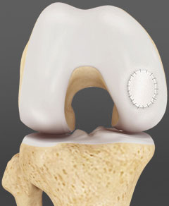
Osteoarticular transfer system (OATS): Osteoarticular transfer system (OATS) is a surgical procedure to treat isolated cartilage defects that are usually 10 to 20 mm in size. The procedure involves the transfer of cartilage plugs taken from non-weight bearing areas of the joint and transferring them into the damaged areas of the joint.
This procedure is not indicated for wide spread damage of cartilage as seen in osteoarthritis.
The procedure is usually performed through arthroscopy. During the procedure, the plugs taken are usually larger and therefore only one or two plugs are needed to fill the area of cartilage damage. The area of damaged cartilage is prepared using a coring tool which makes a perfectly round hole in the bone in damage. The hole is drilled to a size that fits the plug. Next, the plug of the normal cartilage is harvested from a non-weight bearing area of the knee and is then implanted into the hole that was created in the damaged area. The size of the plug used should be slightly larger than the hole so that it fits into the position. This procedure allows the newly implanted bone and cartilage to grow in the defected area.
Possible complications of OATS include donor site morbidity causing pain, avascular necrosis, and fracture. Other complications such as hemarthrosis, effusion and pain may also occur. Following OATS rehabilitation is recommended with the use of crutches and limiting the range of motion.